Bacterial endospores are among the most resilient forms of life on earth and are intrinsically resistant to extreme environments and antimicrobial treatments. Their resilience is explained by unique cellular structures formed by a complex developmental process often initiated in response to nutrient deprivation. Although the macromolecular structures of spores from different bacterial species are similar, their resistance to environmental insults differs widely. It is not known which of the factors attributed to spore resistance confer very high-level heat resistance. Here, we provide conclusive evidence that in Bacillus subtilis, this is due to the presence of a mobile genetic element (Tn1546-like) carrying five predicted operons, one of which contains genes that encode homologs of SpoVAC, SpoVAD and SpoVAEb and four other genes encoding proteins with unknown functions. This operon, named spoVA2mob, confers high-level heat resistance to spores. Deletion of spoVA2mobin a B. subtilis strain carrying Tn1546 renders heat-sensitive spores while transfer of spoVA2mobinto B. subtilis 168 yields highly heat-resistant spores. On the basis of the genetic conservation of different spoVA operons among spore-forming species of Bacillaceae, we propose an evolutionary scenario for the emergence of extremely heat-resistant spores in B. subtilis, B. licheniformis and B. amyloliquefaciens. This discovery opens up avenues for improved detection and control of spore-forming bacteria able to produce highly heat-resistant spores.
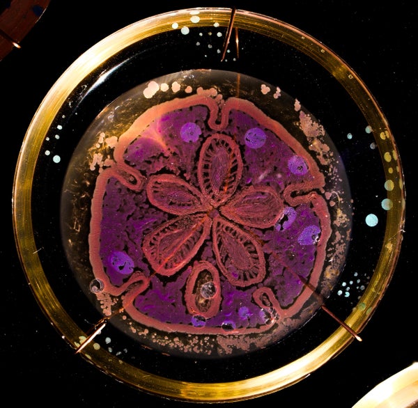
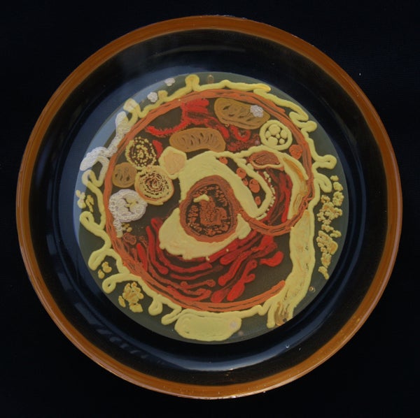
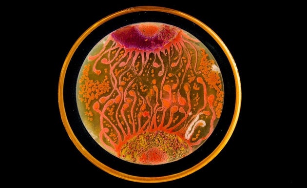
Peñil’s piece “Cell to Cell,” which won the People’s Choice award in the 2015 ASM Agar Art contest.
You’ve never seen bacteria quite like this before.
Mixed media artistMaria Peñil Cobo, who was born in Spain and currently resides in Massachusetts, told The Huffington Post on Thursday that she has often turned to nature as inspiration for her artwork. But instead of looking to vast oceans or forest landscapes, it’s the muchsmaller ecosystems that fascinate her the most.
Peñil has spent the past five years growing colorful bacteria, with help from microbiologist Dr. Mehmet Berkmen, and then “painting” the microbes into stunning masterpieces.
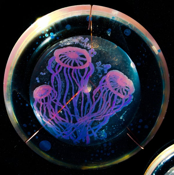
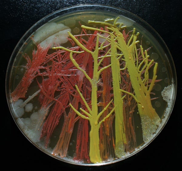
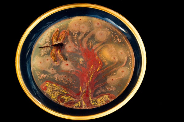
MARIA PEñIL COBO/MEHMET BERKMENPeñil’s “Neurons,” which won first place in the 2015 ASM Agar Art contest.
“It is very technically difficult,” Berkmen, a staff scientist at the Ipswich, Massachusetts-based companyNew England Biolabs, told HuffPost. “You have to imagine that these bacteria we’re using are all different species. ... Each one grows differently and eats differently. Some don’t become colorful immediately, while others become old and then get their color.”
Berkmen taught Peñil how to “paint” with bacteria on agar, a gelatinous substance in which jungles of bacteria can grow. The artist uses a petri dish as her canvas.
Now, watch as the bacteria grow below.
So far, Peñil has attempted to “paint” with bacteria found on her own lips — which she collected after kissing a petri dish — as well as the germs that grew when she put her own house key on the dish.
Peñil, who will begiving a TED Talk about her workin Chicago on April 9, said that she hopes her artwork will shift the public dialogue around bacteria from one of fear and disgust to one of appreciation and curiosity.
“I’m a scientist, and I appreciate this project a lot,” Berkmen said. “When we do science, there is always an element of art, and while Maria is doing pure art, there is an element of science in what we are observing. We are observing scientific phenomena.”
Scroll down to see more of Peñil’s bacteria artwork below.
A protein fragment released by filaments of the fungusCandida albicansdestroys host cells. This is the first demonstration that human fungal pathogens other than moulds can release toxic peptides.
Subject terms:
The fungusCandida albicans, a common cause of infection in mucosal tissues, forms long filaments called hyphae, comprised of tubular cells, that are required for virulence in animals. Hyphal-associated adhesion proteins bind to host tissue, which is then degraded by hydrolytic enzymes. However, until now, no hyphal toxin that damages host cells has been identified. Because of this, the fungus is considered to be an 'accidental' pathogen that benignly inhabits our mucosal tissues, causing tissue damage by happenstance rather than design. That view must now change drastically. Moyeset al.reveal a mechanism through which hyphae actively wage war on our cells — the production of a toxin that the authors call Candidalysin.
Interactions betweenC. albicansand epithelial cells, which line the body's cavities, occur during the early stages of mucosal infections such as thrush and vaginitis. Hyphae elicitseveral epithelial-cell responses, including the production of signalling molecules called cytokines that recruit cells of the immune system to defend tissues, and loss of cell integrity through cell-membrane deterioration. Moyeset al. discovered that a strain ofC. albicansin which the geneECE1was mutated could not elicit epithelial-cell responses, despite growing apparently normal hyphae. Moreover, the authors validated these tissue-culture observationsin vivo— theECE1mutant was unable to reliably infect mucosa in a zebrafish swimbladder model and a mouse model of thrush.
ECE1was one of the first genes to be identified in hyphal-specific expression screens more than 20 years ago, yet until now it has been one of the most poorly understood genes inC. albicans. In fact,ECE1is among the most highly expressed genes in hyphae, but its role has not previously been investigated thoroughly because mutants show no defects in hyphal morphology or cell proliferation. Thus, the function of the Ece1 protein has remained a puzzle.
How does Ece1 promote epithelial-cell responses? The protein's amino-acid sequence suggests that it is secreted from hyphae as a group of eight short protein fragments, or peptides, and so would be well positioned to interact with host cells. Moyes and colleagues confirmed that all eight Ece1 peptides are secreted from hyphae. Analysis of synthetic versions of each peptide revealed that one, Ece1-III, elicits the same responses from epithelial cells as do hyphae. Moreover, precise deletion of the genetic region that codes for only Ece1-III created a mutantC. albicansthat secreted the remaining seven peptides, but did not elicit epithelial-cell responses or cause mucosal disease in animal models. These results clearly demonstrate that Ece1-III mediates the pathogenic activity associated withECE1.
By what mechanism does Ece1-III exert this activity? Certain chemical and structural features indicate that Ece1-III might function like peptide toxins, such as the bee-venom toxin melittin. Indeed, the authors show that the peptide causes rapid and transient permeabilization of artificial cell membranesin vitro. These activities are enhanced in the presence of cholesterol, a component of animal — but not fungal — membranes. The researchers conclude that Ece1-III acts as a peptide toxin, which they name Candidalysin.
Moyes and colleagues' study establishes thatC. albicanshyphae evolved to damage host cells. When combined with our knowledge of hyphal adhesin proteins and enzymes, a simple program of tissue destruction emerges (Fig. 1). First, the hyphal-specific adhesin Hwp1 attaches to mucosal surfaces. Second, the hyphal-specific invasion protein Als3, acting with the protein Ssa1, binds to receptors on the surface of the host cell, promoting engulfment of the hypha by the host cell8. Finally, Candidalysin accumulates in the invasion pocket around the hypha, attacking the host's cholesterol-containing membrane.
Figure 1: A toxic relationship.
The pathogenic fungusCandida albicansinfects its host by forming filamentous structures called hyphae. Proteins on the hyphal surface — the adhesin Hwp1, the invasin Als3 and its partner Ssa1 — make contact with the host cell directly or through receptor proteins to promote adhesion and engulfment of the hypha by the host. Moyeset al.2report that the protein Ece1 is secreted from the hypha as eight short peptides. One of these, Candidalysin, acts as a toxin that accumulates in the invasion pocket and attacks the host-cell membrane, leading to membrane permeabilization and the induction of host defences.
This attack leads to membrane permeabilization, leakage of cell contents and a defensive cytokine response, which serves to limit the size of theC. albicanspopulation in healthy individuals. However, impaired defences in people with conditions such as AIDS, diabetes and some cancers permitC. albicansgrowth and consequent disease.
We used whole-genome design and complete chemical synthesis to minimize the 1079–kilobase pair synthetic genome of Mycoplasma mycoides JCVI-syn1.0. An initial design, based on collective knowledge of molecular biology combined with limited transposon mutagenesis data, failed to produce a viable cell. Improved transposon mutagenesis methods revealed a class of quasi-essential genes that are needed for robust growth, explaining the failure of our initial design. Three cycles of design, synthesis, and testing, with retention of quasi-essential genes, produced JCVI-syn3.0 (531 kilobase pairs, 473 genes), which has a genome smaller than that of any autonomously replicating cell found in nature. JCVI-syn3.0 retains almost all genes involved in the synthesis and processing of macromolecules. Unexpectedly, it also contains 149 genes with unknown biological functions. JCVI-syn3.0 is a versatile platform for investigating the core functions of life and for exploring whole-genome design.
Cite this article as C. A. Hutchison III et al., Science 351, aad6253 (2016). DOI: 10.1126/science.aad6253
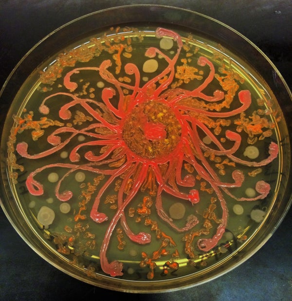
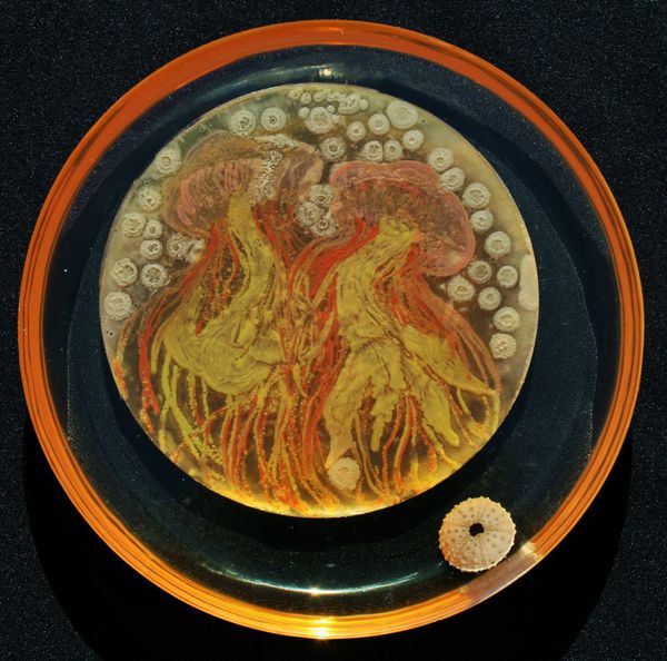
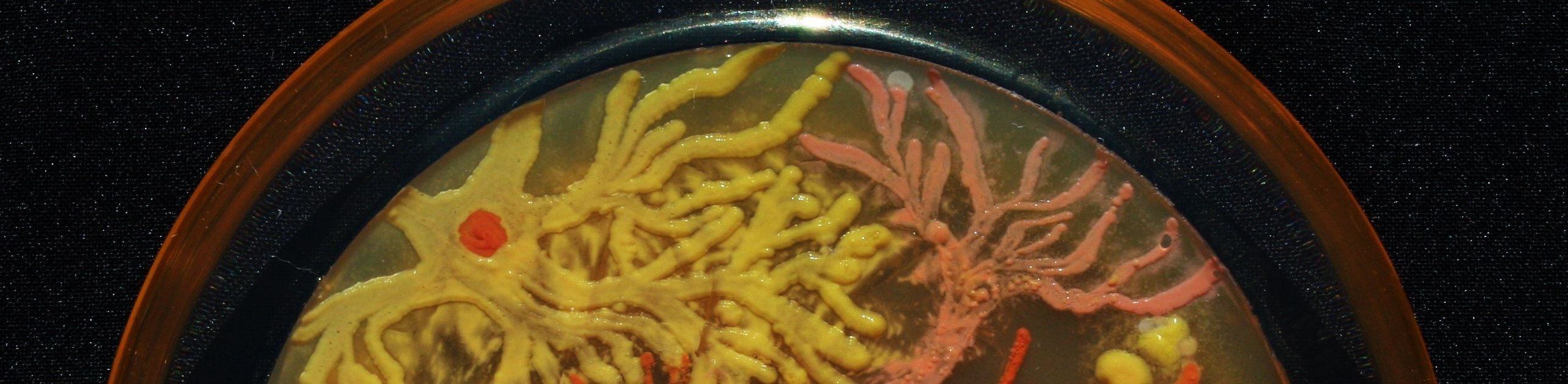 MARIA PEñIL COBO/MEHMET BERKMEN
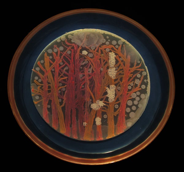