Reloj

jueves, 25 de septiembre de 2014

A faster Rubisco with potential to increase photosynthesis in crops

In photosynthetic organisms, d-ribulose-1,5-bisphosphate carboxylase/oxygenase (Rubisco) is the major enzyme assimilating atmospheric CO2 into the biosphere1. Owing to the wasteful oxygenase activity and slow turnover of Rubisco, the enzyme is among the most important targets for improving the photosynthetic efficiency of vascular plants2, 3. It has been anticipated that introducing the CO2-concentrating mechanism (CCM) from cyanobacteria into plants could enhance crop yield4, 5, 6. However, the complex nature of Rubisco’s assembly has made manipulation of the enzyme extremely challenging, and attempts to replace it in plants with the enzymes from cyanobacteria and red algae have not been successful7, 8. Here we report two transplastomic tobacco lines with functional Rubisco from the cyanobacterium Synechococcus elongatus PCC7942 (Se7942). We knocked out the native tobacco gene encoding the large subunit of Rubisco by inserting the large and small subunit genes of the Se7942 enzyme, in combination with either the corresponding Se7942 assembly chaperone, RbcX, or an internal carboxysomal protein, CcmM35, which incorporates three small subunit-like domains9, 10. Se7942 Rubisco and CcmM35 formed macromolecular complexes within the chloroplast stroma, mirroring an early step in the biogenesis of cyanobacterial β-carboxysomes11, 12. Both transformed lines were photosynthetically competent, supporting autotrophic growth, and their respective forms of Rubisco had higher rates of CO2 fixation per unit of enzyme than the tobacco control. These transplastomic tobacco lines represent an important step towards improved photosynthesis in plants and will be valuable hosts for future addition of the remaining components of the cyanobacterial CCM, such as inorganic carbon transporters and the β-carboxysome shell proteins4, 5, 6.

Phenotype of the wild-type and transplastomic tobacco lines.


Enlace al trabajo

lunes, 2 de junio de 2014

Retroviruses, the Placenta, and the Genomic Junk Drawer


Tomado de Small Things Considered
Figure1 Figure 1. The ubiquitous junk drawer, filled with items that might come in handy one day. Source.
At our very core, we are all hoarders
By now, many of us are aware that a considerable portion (45% or more) of the human genome consists of transposable elements. These are mobile genetic sequences, such as Alu repeats and long and short interspersed nuclear elements (LINEs and SINEs). A whopping 18% of this so-called "dark matter of the genome" is retroviral sequences left over from ancient infections of germ line cells. This means that, in total, ~8% of the human genome is retroviral, compared to only ~1.5% that codes for human genes — a situation that led my Ph.D. advisor to claim that there are more retroviruses in us than there is us in us! Due to mutations, the viruses no longer function as viruses, but are a collection of broken parts.
Considering that the human body carries out ~10,000 billion cell divisions in a lifetime, the replication and maintenance of this viral junk heap requires a considerable amount of energy and resources. So why do we keep it around? Probably for the very reason that nearly every household has a junk drawer (Figure 1): weak purifying selection (read, lack of motivation to clean up), combined with the hunch that “this stuff might come in handy someday.” In fact, it appears that an important evolutionary innovation — the placenta — has depended on parts pulled from the “genomic junk drawer.”
Figure2 Figure 2. The invasion of the uterine wall by proliferating trophoblast cells post-fertilization. Source.
The birth of the placenta
The placenta evolved in the ancestors of mammals ~150 million years ago, and is a feature of the vast majority of modern mammals, including ourselves. The advantages conferred by this organ are obvious — it protects the developing embryo inside the mother, shields it from the elements, and ensures a steady supply of nutrients, moisture, and the appropriate temperature for growth and comfortable napping. How did this lucky innovation come about?
Some hints lie in the details of placental development, which begins five days after fertilization with the formation of a clump of cells called the blastocyst (Figure 2). The blastocyst consists of an inner cell mass that will become the embryo, and an outer layer of cells called trophoblasts, from which the placenta arises. The trophoblasts invade the uterine wall and proliferate, in a tumor-like manner, differentiating into a specialized cell type, the cytotrophoblasts. The cytotrophoblasts then fuse together into a layer of multinucleated cells, the syncytiotrophoblasts, creating a sac firmly attached to the uterine wall. This layer of fused cells creates a specialized barrier between mother and fetus, through which the fetus can acquire nutrients and growth hormones while ridding itself of waste products. The fetus will express both maternal and paternal proteins. While the former will be tolerated by the mother’s immune system, the latter will be recognized as invasive. It is the placenta that protects the fetus from attack by the mother’s immune system.
The italicized portions above have suggested an uncomfortable comparison of pregnancy to invasion by a parasite, with the mother’s physiology being reworked to serve the needs of the fetus. Whether you like the analogy or not, the placenta's position at the materno-fetal interface indeed likens it to an interface between a pathogen and its host. This situation requires an elevated level of adaptability — a challenge for complex DNA organisms like us, with our stodgy mutation rates, but typical of faster-replicating entities such as, say, the viruses.
If it walks like a retrovirus...
The layer of fused cells that forms the membrane between mother and fetus is the key histological feature of the placenta. Cell-cell fusion is not a common feature of mammalian cells, but it is the basis of how retroviruses and other enveloped viruses enter host cells during an infection. Here’s the story: the retroviral membrane is studded with proteins designated “Env” for envelope that are made up of two parts. part SU) for surface) is responsible for binding to a specific receptor on the host cell, while the other part (TM, for transmembrane) fuses the cell and virus membranes together, so that the virus can enter the cell. By the same mechanism, a cell expressing Env can fuse with another cell expressing the appropriate receptor protein. Due to this ability, the retroviral Env protein is said to be "fusogenic."
By the time the placenta evolved there were already many retroviruses around that had integrated into the genomes of vertebrate species (so-called "endogenous retroviruses" or "ERVs"). Once they've inserted into the genome, ERVs evolve neutrally and in time become inactivated by the accumulation of mutations and by mechanisms that the host uses to repress their expression. Env coding regions, however, are sometimes conserved after integration. The thinking is that the Env proteins can block receptors from being used by any retroviruses that use the same receptor. Therefore, if a recently integrated retrovirus is defective for replication but can still express Env, it can confer protection on the host from infection with similar retroviruses. In other words, Env coding regions, like stray rubber bands in a junk drawer, are always in supply.
Figure3 Figure 3. Retrovirus particles budding from rhesus macaque placenta cells. Source.
Clues that mammals actually do avail themselves of these spare parts emerged in the 1970’s: Electron micrographs of placental tissue from humans and a variety of other animals showed retrovirus-like particles budding from placental cells, particularly within the syncytiotrophoblast layer — the layer consisting of fused cells (Figure 2). The presence of these particles in healthy tissues of many placental mammals prompted a few brave researchers to entertain the idea that ERVs actually play an essential role in placental development. The rather astounding implication was that an accidental infection of a mammalian ancestor by a retrovirus was responsible for the evolution of a novel mechanism of reproduction, one that launched the entire lineage of placental mammals no less.
Within a few years, a couple of prospects for the putative placental retrovirus were found among the HERV-W and HERV-FRD families of human ERVs (HERV stands for human ERV). Comparative genomics reveals that HERV-W infected the germ line of an ancestor of the great apes (including humans) over 25 million years ago (mya). HERV-FRD is even older, having first infected the ancestors of all monkeys except prosimians some 40 mya. Although during their long tenure in the genome both retroviruses have accumulated mutations that render them incapable of producing infectious virions, the reading frame across the envelope genes (env) are still intact. Additionally, they fulfill the three requirements for a gene to be involved in placental formation: 1) placenta-specific expression, 2) fusogenicity, and 3) sequence conservation across multiple species, which suggests an essential role in survival. In situ studies reveal that HERV-W is expressed in many placental cell types, whereas HERV-FRD expression is limited to a particular type of cytotrophoblasts. In experiments on primary cultures of cytotrophoblasts, inhibiting either Env—but especially that of HERV-FRD—reduces the fusion efficiency and the production of the pregnancy-hormone human chorionic gonadotropin. It sure sounds like both HERV-W and HERV-FRD contribute to placental development in humans. HERV-W was named syncytin-1 and HERV-FRD, syncytin-2 for their ability to fuse cells and form syncytia.
Once they knew what to look for, researchers made fast work of screening the mouse genome for ERVs with open reading frames across the env gene. They found two sequences, now known as syncytin-A and –B (the nomenclature changes from 1 and 2 in humans to A and B in mice), which met the three criteria for bona fide syncytinhood, mentioned above. Furthermore, when researchers prevented expression of syncytin-A in mice, unfused cells accumulated, which disrupted the syncytiotrophoblast layer and caused the embryos to die at mid-gestation. This finding constituted proof that a gene of retroviral origin is essential for survival in mice. Prevention of syncytin-B expression resulted in impaired formation of one of the two placental layers found in mice, confirming that it, too, had a role in placentation.
Remarkably, mouse syncytin-A and -B differ from primate syncytin-1 and -2 in key respects — they are not in analogous locations on the chromosomes and are from distinct retrovirus families. In other words, they represent separate, independent gene capture events (as if the mouse and primate lineages each pulled different rubber bands from the junk drawer and used them for the same purpose). An amazing pattern of convergent evolution has emerged as more examples of independently acquired syncytins have surfaced in various mammalian orders: syncytin-Car1 is found in carnivores; syncytin-Ory1 in lagomorphs (hares, rabbits, and pikas); syncytin-Rum1 in cows and various other ruminants (Figure 4a); and most recently, syncytin-Mar1 in squirrels.
Figure4 Figure 4. a) Various retrovirus env genes have been captured independently by different lineages of placental mammals. Purple triangles represent env capture events. (b) Diversity of placental structures might be explained by use of different retrovirus env genes (syncytins). Source.
Rummaging through the genomic junk drawer
Because ERVs can cause disease in a number of ways, the host generally represses their transcription. Repression is particularly strict in embryonic stem cells, where unchecked ERV activity could interfere with the complex and delicately timed pathways required during development. In stark contrast, repression is relaxed in the progenitors of placental cells, and transcripts from multiple ERV families are produced. Why would that be? The placenta is a transient organ, fated to be eaten or plunked into a specimen pan on the way to the trash. At the same time, the placenta's position at the materno-fetal interface requires an elevated level of adaptability. The derepression of ERVs might be seen as a means of harnessing a little b of the adaptive potential of faster-mutating entities in a context where costs to the host are not too high, due to the transient nature of the placenta.
The marked diversity of placental structures among mammalian species (Figure 4b) may be an outcome of this ERV-induced plasticity, as hypothesized by members of the Heidmann lab, who are leading the charge in identifying and characterizing syncytins. Since the various syncytins originated from different retroviruses, they exhibit differences in expression levels, fusogenicity, and receptor usage. Such differences likely impact placental structure. Also reflecting this plasticity is evidence within some species that older syncytin genes are phased out when a new env sequence is captured (Figure 4a, Rodentia and Haplorrhini orders). This ongoing replacement model would explain the discrepancy between the ages of the known syncytins (captured between 12 and 60 mya) and the emergence of a primitive placenta some 150 mya.
All of this suggests the oh-so-elegant junk drawer model: Derepressing ERV expression in placental progenitor cells can be likened to opening the junk drawer and rummaging around for anything that might be of use. Since ERVs are not random bits of DNA but complex sequences that code for various functional domains such as binding sites, proteases, promoters, and fusion domains, the likelihood of pulling something useful out of the ERV junk drawer is rather high, even if the item wasn't originally designed for the particular task. It is amazing to think that an evolutionary innovation of such consequence — the placenta — was essentially pulled from the genomic junk drawer!

miércoles, 28 de mayo de 2014

A draft map of the human proteome

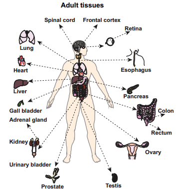
The availability of human genome sequence has transformed biomedical research over the past decade. However, an equivalent map for the human proteome with direct measurements of proteins and peptides does not exist yet. Here we present a draft map of the human proteome using high-resolution Fourier-transform mass spectrometry. In-depth proteomic profiling of 30 histologically normal human samples, including 17 adult tissues, 7 fetal tissues and 6 purified primary haematopoietic cells, resulted in identification of proteins encoded by 17,294 genes accounting for approximately 84% of the total annotated protein-coding genes in humans. A unique and comprehensive strategy for proteogenomic analysis enabled us to discover a number of novel protein-coding regions, which includes translated pseudogenes, non-coding RNAs and upstream open reading frames. This large human proteome catalogue (available as an interactive web-based resource at http://www.humanproteomemap.org) will complement available human genome and transcriptome data to accelerate biomedical research in health and disease.


Acceso al trabajo de Nature
Acceso al segundo trabajo de Nature

jueves, 8 de mayo de 2014

A semi-synthetic organism with an expanded genetic alphabet

Triphosphates of hydrophobic nucleotides d5SICS and dNaM are imported into Escherichia coli by an exogenous algal nucleotide triphosphate transporter and then used by an endogenous polymerase to replicate, and faithfully maintain over many generations of growth, a plasmid containing the d5SICS–dNaM unnatural base pair. Neither the presence of the unnatural triphosphates nor the replication of the UBP introduces a notable growth burden. Lastly, we find that the UBP is not efficiently excised by DNA repair pathways. Thus, the resulting bacterium is the first organism to propagate stably an expanded genetic alphabetNucleoside triphosphate stability and import.

a, Chemical structure of the d5SICS–dNaM UBP compared to the natural dG–dC base pair. b, Composition analysis of d5SICS and dNaM in the media (top) and cytoplasmic (bottom) fractions of cells expressing PtNTT2 after 30 min incubation; dA shown for comparison. 3P, 2P, 1P and 0P correspond to triphosphate, diphosphate, monophosphate and nucleoside, respectively; [3P] is the intracellular concentration of triphosphate. Error bars represent s.d. of the mean, n = 3.

Enlace al trabajo

viernes, 2 de mayo de 2014

El vinagre, un nuevo antiséptico?


Mycobacteria are best known for causing tuberculosis and leprosy, but infections with nontuberculous mycobacteria are an increasing problem after surgical or cosmetic procedures or in the lungs of cystic fibrosis and immunosuppressed patients. Killing mycobacteria is important because Mycobacterium tuberculosis strains can be multidrug resistant and therefore potentially fatal biohazards, and environmental mycobacteria must be thoroughly eliminated from surgical implements and respiratory equipment. Currently used mycobactericidal disinfectants can be toxic, unstable, and expensive. We fortuitously found that acetic acid kills mycobacteria and then showed that it is an effective mycobactericidal agent, even against the very resistant, clinically important Mycobacterium abscessus complex. Vinegar has been used for thousands of years as a common disinfectant, and if it can kill mycobacteria, the most disinfectant-resistant bacteria, it may prove to be a broadly effective, economical biocide with potential usefulness in health care settings and laboratories, especially in resource-poor countries.

Enlace al trabajo científico

jueves, 27 de marzo de 2014

First synthetic yeast chromosome revealed


As a eukaryote, a category that includes humans and other animals, S. cerevisiae has a more complex genome than Venter's parasite. The synthetic yeast chromosome — which has been stripped of some DNA sequences and other elements — is 272,871 base pairs long, representing about 2.5% of the 12-million-base-pair S. cerevisiae genome.The researchers, who report their accomplishment in Science1, have formed an international consortium to create a synthetic version of the full S. cerevisiae genome within 5 years.


Enlace a la noticia


Total Synthesis of a Functional Designer Eukaryotic Chromosome


Rapid advances in DNA synthesis techniques have made it possible to engineer viruses, biochemical pathways and assemble bacterial genomes. Here, we report the synthesis of a functional 272,871–base pair designer eukaryotic chromosome, synIII, which is based on the 316,617–base pair native Saccharomyces cerevisiae chromosome III. Changes to synIII include TAG/TAA stop-codon replacements, deletion of subtelomeric regions, introns, transfer RNAs, transposons, and silent mating loci as well as insertion of loxPsym sites to enable genome scrambling. SynIII is functional in S. cerevisiae. Scrambling of the chromosome in a heterozygous diploid reveals a large increase in a-mater derivatives resulting from loss of the MATα allele on synIII. The complete design and synthesis of synIII establishes S. cerevisiae as the basis for designer eukaryotic genome biology.

Enlace al paper de Science                    

Bacterial solutions to multicellularity: a tale of biofilms, filaments and fruiting bodies

Although bacteria frequently live as unicellular organisms, many spend at least part of their lives in complex communities, and some have adopted truly multicellular lifestyles and have abandoned unicellular growth. These transitions to multicellularity have occurred independently several times for various ecological reasons, resulting in a broad range of phenotypes. In this Review, we discuss the strategies that are used by bacteria to form and grow in multicellular structures that have hallmark features of multicellularity, including morphological differentiation, programmed cell death and patterning. In addition, we examine the evolutionary and ecological factors that lead to the wide range of coordinated multicellular behaviours that are observed in bacteria.


Enlace al artículo

El corazón púrpura de la fotosíntesis

The structure of a photosynthetic complex from a purple bacterium reveals a new class of light-harvesting protein and the channels that might allow electron-transporting molecules to escape this otherwise closed system.

Typical organization of a photosynthetic unit in the intracellular membrane of purple bacteria.

Light-harvesting complex 2 (LH2) contains two groups of bacteriochlorophyll (BChL) pigments, B800 and B850. Light energy absorbed by B800 is transferred to B850 (blue arrows). It is then passed to another group of BChLs (B880) in light-harvesting complex 1 (LH1), and finally into the reaction centre (RC). The excited RC fully reduces ubiquinone molecules (Q), which leave the LH1–RC complex and pass into the membrane, transferring electrons (e) to the cytochrome bc1 complex; this transfer forms part of a cyclic electron-transport pathway (red arrows) that drives photosynthesis. This pathway is completed by a soluble protein, cytochrome c2 (Cyt c2). The periplasm is the region between the cell membrane and the cell wall of the bacterium.


domingo, 23 de marzo de 2014

Structural Studies of Planctomycete Gemmata obscuriglobus Support Cell Compartmentalisation in a Bacterium.

Members of phylum Planctomycetes have been proposed to possess atypical cell organisation for the Bacteria, having a structure of sectioned cells consistent with internal compartments surrounded by membranes. Here via electron tomography we confirm the presence of compartments in the planctomycete Gemmata obscuriglobus cells. Resulting 3-D models for the most prominent structures, nuclear body and riboplasm, demonstrate their entirely membrane - enclosed nature. Immunogold localization of the FtsK protein also supports the internal organisation of G.obscuriglobus cells and their unique mechanism of cell division. We discuss how these new data expand our knowledge on bacterial cell biology and suggest evolutionary consequences of the findings.

Enlace al trabajo

Taking aim at wall teichoic acid synthesis: new biology and new leads for antibiotics.

Wall teichoic acids are a major and integral component of the Gram-positive cell wall. These structures are present across all species of Gram-positive bacteria and constitute roughly half of the cell wall. Despite decades of careful investigation, a definitive physiological function for wall teichoic acids remains elusive. Advances in the genetics and biochemistry of wall teichoic acid synthesis have led to a new understanding of the complexity of cell wall synthesis in Gram-positive bacteria. Indeed, these innovations have provided new molecular tools available to probe the synthesis and function of these cell wall structures. Among recent discoveries are unexpected roles for wall teichoic acid in cell division, coordination of peptidoglycan synthesis and β-lactam resistance in methicillin-resistant Staphylococcus aureus (MRSA). Notably, wall teichoic acid biogenesis has emerged as a bona fide drug target in S. aureus, where remarkable synthetic-viable interactions among biosynthetic genes have been leveraged for the discovery and characterization of novel inhibitors of the pathway.


 Enlace al trabajo

jueves, 13 de marzo de 2014

SecA is required for membrane targeting of the cell division protein DivIVA in vivo


The conserved protein DivIVA is involved in different morphogenetic processes in Gram-positive bacteria. In Bacillus subtilis, the protein localizes to the cell division site and cell poles, and functions as a scaffold for proteins that regulate division site selection, and for proteins that are required for sporulation. To identify other proteins that bind to DivIVA, we performed an in vivo cross-linking experiment. A possible candidate that emerged was the secretion motor ATPase SecA. SecA mutants have been described that inhibit sporulation, and since DivIVA is necessary for sporulation, we examined the localization of DivIVA in these mutants. Surprisingly, DivIVA was delocalized, suggesting that SecA is required for DivIVA targeting. To further corroborate this, we performed SecA depletion and inhibition experiments, which provided further indications that DivIVA localization depends on SecA. Cell fractionation experiments showed that SecA is important for binding of DivIVA to the cell membrane. This was unexpected since DivIVA does not contain a signal sequence, and is able to bind to artificial lipid membranes in vitro without support of other proteins. SecA is required for protein secretion and membrane insertion, and therefore its role in DivIVA localization is likely indirect. Possible alternative roles of SecA in DivIVA folding and/or targeting are discussed.

FIGURE 3

 Control experiments to confirm the cross-linking of SecA with DivIVA. (A) Pull-down experiment with His-DivIVA, DivIVA-GFP-His and GFP-His as bait proteins (strains BSN50, 3308 and BSN73, respectively). Pull-down fractions were analyzed by Western blotting for the presence of SecA (upper panel). To demonstrate that the bait proteins were pulled down efficiently, fractions were analyzed by Western blotting using DivIVA (middle panel) and GFP antisera (lower panel). (B) Pull-down experiment using SecA-His and GFP-His as bait proteins (strains BSN131, and 4097, respectively). Protein fractions were separated by SDS-PAGE (upper panel), and the presence of DivIVA was analyzed by Western blotting (lower panel). SecY, a known interaction partner of SecA, was pulled down with SecA-His. The protein band at around 55 kDa is elongation factor Tu (TufA), which appears to be an unspecific by-catch. The theoretical position of DivIVA on the SDS PAGE gel is indicated in brackets.

Enlace al trabajo

martes, 11 de marzo de 2014

Ancient Life in the Information Age


All known organisms share a number of fundamental features that, taken together, point to a common evolutionary history: DNA as the chief molecule of genetic inheritance, proteins as the primary functional molecules, and RNA as an informational intermediate between the two. The simplest explanation for why organisms share these common features is that they are inherited from a last universal common ancestor (LUCA), which sits at the root of the tree of life. Most studies of gene duplications that occurred prior to the first branch on the tree place LUCA in between the Bacteria and the common ancestor of the Archaea and Eukarya, the three taxonomic domains of cellular life.

The availability of the genome sequences from so many species across the tree of life has made it possible to look for common genomic traits that were most likely inherited from LUCA.

Some studies have estimated there to be fewer than 100 LUCA-derived gene families, while others count more than 1,000, depending on how conservatively the methods rule out genes on suspicion of horizontal gene transfer or how liberally they include genes that appear to have been present in LUCA, but subsequently lost. Despite the conflicting results, the new data are yielding insight into ancient life on Earth.

The majority of ancient gene families identified in almost all of these studies are involved in the translation of genetic information into proteins. These ancient gene families represent a range of translation functions, from regulation to ribosomal components. The genetic code at the core of translation is also highly conserved across life. In all likelihood, the enzymes responsible for establishing the genetic code by attaching amino acids to particular tRNAs evolved prior to the time of LUCA, although their evolutionary histories are obscured by subsequent horizontal gene transfers between bacteria and archaea. These results depict a translation system in LUCA that was probably similar to and as sophisticated as those of organisms alive today.

In contrast, few genes involved in the synthesis of DNA are conserved across the tree of life. The enzymes responsible for making deoxyribonucleotides from ribonucleotides exist in three distinct families that only show a weak signature of common descent in their active sites. The only DNA polymerase enzymes that are common across the evolutionary tree are those involved in repair, not the polymerases presently responsible for copying complete chromosomes. RNA polymerases from bacteria, archaea, and eukaryotes, on the other hand, do appear to have been inherited from LUCA, and may have previously functioned as DNA polymerases as well. Taken together, these observations suggest that DNA genomes replaced a genome composed of RNA just prior to or perhaps just after the time of LUCA.

The variety of metabolic strategies observed in modern organisms demonstrates that metabolism is generally less highly conserved, which makes it harder to identify those metabolic pathways that were present in LUCA. Still, various databases organize enzymatic data into metabolic maps, which can be used to uncover highly conserved components of modern metabolic pathways. For example, a recent study combined these data with evolutionary trees of carbon-fixation genes and found that the ancestral carbon-fixation pathway was most likely an amalgam of components currently found in two separate pathways in extant archaea and bacteria: the reductive acetyl-CoA pathway and the reductive citric acid cycle (PLOS Comput Biol, 8:e1002455, 2012). Another taxonomically broad comparison study, focused on amino acid metabolism, uncovered conserved biosynthetic pathways for 8 of the 20 canonical amino acids, and conserved enzymes from pathways for another eight (Genome Biology, 9:R95, 2008).

Finally, LUCA most likely had a phospholipid membrane that set the boundaries between organisms and offered protection from the external environment. The universal presence of genes responsible for targeting proteins to membranes suggests that LUCA’s membrane was replete with proteins. Furthermore, the ubiquity of both catalytic subunits of the membrane-bound ATPase motor also implies that this membrane was impermeable enough to ions that it could be used to generate the proton gradients used by the motor to convert ADP to ATP.

While this detailed understanding of LUCA is relatively recent, Darwin proposed the idea of an early common ancestor to all life in the first edition of Origin of Species, where he wrote, “Therefore I should infer from analogy that probably all the organic beings which have ever lived on this earth have descended from some one primordial form, into which life was first breathed.” Although Darwin’s insight is brilliant for its time, the modern view shows that LUCA is not this “primordial form,” but rather a sophisticated cellular organism that, if alive today, would probably be difficult to distinguish from other extant bacteria or archaea. This means that a great detail of evolution must have taken place between the time of the origin of life and the appearance of LUCA. Continuing advances in evolutionary biology, bioinformatics, and computational biology will give us the tools to describe LUCA and the evolutionary transitions preceding it with unprecedented accuracy and detail.

Aaron David Goldman is an assistant professor of biology at Oberlin College. His research employs bioinformatics and systems biology tools to study the genome and metabolism of LUCA and their connections to evolutionary predecessors. 

Enlace al texto

Enlace a LUCApedia


miércoles, 5 de marzo de 2014

The Role of CRISPR-Cas Systems in Virulence of Pathogenic Bacteria

Clustered regularly interspaced short palindromic repeats (CRISPR) and CRISPR-associated (Cas) genes are present in many bacterial and archaeal genomes. Since the discovery of the typical CRISPR loci in the 1980s, well before their physiological role was revealed, their variable sequences have been used as a complementary typing tool in diagnostic, epidemiologic, and evolutionary analyses of prokaryotic strains. The discovery that CRISPR spacers are often identical to sequence fragments of mobile genetic elements was a major breakthrough that eventually led to the elucidation of CRISPR-Cas as an adaptive immunity system. Key elements of this unique prokaryotic defense system are small CRISPR RNAs that guide nucleases to complementary target nucleic acids of invading viruses and plasmids, generally followed by the degradation of the invader. In addition, several recent studies have pointed at direct links of CRISPR-Cas to regulation of a range of stress-related phenomena. An interesting example concerns a pathogenic bacterium that possesses a CRISPR-associated ribonucleoprotein complex that may play a dual role in defense and/or virulence. In this review, we describe recently reported cases of potential involvement of CRISPR-Cas systems in bacterial stress responses in general and bacterial virulence in particular.

Enlace al trabajo

Sulphoglycolysis in Escherichia coli K-12 closes a gap in the biogeochemical sulphur cycle

Sulphoquinovose (SQ, 6-deoxy-6-sulphoglucose) has been known for 50 years as the polar headgroup of the plant sulpholipid in the photosynthetic membranes of all higher plants, mosses, ferns, algae and most photosynthetic bacteria. It is also found in some non-photosynthetic bacteria, and SQ is part of the surface layer of some Archaea. The estimated annual production of SQ is 10,000,000,000 tonnes (10 petagrams), thus it comprises a major portion of the organo-sulphur in nature, where SQ is degraded by bacteria. However, despite evidence for at least three different degradative pathways in bacteria, no enzymic reaction or gene in any pathway has been defined, although a sulphoglycolytic pathway has been proposed7. Here we show that Escherichia coli K-12, the most widely studied prokaryotic model organism, performs sulphoglycolysis, in addition to standard glycolysis. SQ is catabolised through four newly discovered reactions that we established using purified, heterologously expressed enzymes: SQ isomerase, 6-deoxy-6-sulphofructose (SF) kinase, 6-deoxy-6-sulphofructose-1-phosphate (SFP) aldolase, and 3-sulpholactaldehyde (SLA) reductase. The enzymes are encoded in a ten-gene cluster, which probably also encodes regulation, transport and degradation of the whole sulpholipid; the gene cluster is present in almost all (>91%) available E. coli genomes, and is widespread in Enterobacteriaceae. The pathway yields dihydroxyacetone phosphate (DHAP), which powers energy conservation and growth of E. coli, and the sulphonate product 2,3-dihydroxypropane-1-sulphonate (DHPS), which is excreted. DHPS is mineralized by other bacteria, thus closing the sulphur cycle within a bacterial community.


Enlace al trabajo

Identification of a conserved branched RNA structure that functions as a factor-independent terminator

All cells regulate which regions of DNA are transcribed to RNA. Controlling where transcription terminates is an essential part of this regulation. In bacterial cells, RNA structures, referred to as factor-independent terminators, can interact with RNA polymerase to direct termination. These structures are typically inverted sequence repeats that form an RNA hairpin followed by several uridine residues. We identified a branched RNA structure that functions as a factor-independent terminator. The terminated product is a functional small RNA, but termination is inefficient, allowing transcription of downstream genes. Additional branched terminators are encoded in bacterial chromosomes, demonstrating that this unusual terminator is not unique. This work reveals an unappreciated structural diversity of factor-independent terminators and will inform annotation of bacterial genomes.    

Enlace al trabajo                    

jueves, 27 de febrero de 2014

A new metabolic cell-wall labelling method reveals peptidoglycan in Chlamydia trachomatis

Peptidoglycan (PG), an essential structure in the cell walls of the vast majority of bacteria, is critical for division and maintaining cell shape and hydrostatic pressure. Bacteria comprising the Chlamydiales were thought to be one of the few exceptions. Chlamydia harbour genes for PG biosynthesis and exhibit susceptibility to ‘anti-PG’ antibiotics yet attempts to detect PG in any chlamydial species have proven unsuccessful (the ‘chlamydial anomaly’). We used a novel approach to metabolically label chlamydial PG using d-amino acid dipeptide probes and click chemistry. Replicating Chlamydia trachomatis were labelled with these probes throughout their biphasic developmental life cycle, and the results of differential probe incorporation experiments conducted in the presence of ampicillin are consistent with the presence of chlamydial PG-modifying enzymes. These findings culminate 50 years of speculation and debate concerning the chlamydial anomaly and are the strongest evidence so far that chlamydial species possess functional PG.

Fluorescent labelling of intracellular C. trachomatis PG.

Fig. 1. Fluorescent labelling of intracellular C. trachomatis PG. 
ae, Differential interference contrast (DIC) (a) and fluorescent (be) microscopy of L2 cells infected for 18 h with C. trachomatis in the presence of the dipeptide probe EDA-DA (1mM). Subsequent binding of the probe to an azide modified Alexa Fluor 488 (green) was achieved via click chemistry. Antibody to MOMP (red) was used to label chlamydial EBs and RBs. DAPI (blue) was used for nuclear staining. be show a merge of all three fluorescent channels. Boxes indicate location of chlamydial inclusions, and magnification of the boxes is provided in ce. Fluorescent images are maximum intensity projections of deconvoluted z-stacks. 

Different walls for rods and balls: the diversity of peptidoglycan


Peptidoglycan performs the essential role of resisting turgor in the cell walls of most bacteria. It determines cell shape, and its biosynthesis is the target for many important antibiotics. The fundamental chemical building blocks of peptidoglycan are conserved: repeating disaccharides cross-linked by peptides. However, these blocks come in many varieties and can be assembled in different ways. So beyond the fundamental similarity, prodigious chemical, organizational and architectural diversity is revealed. Here, we track the evolution of our current understanding of peptidoglycan and underpinning technical and methodological developments. The origin and function of chemical diversity is discussed with respect to some well-studied example species. We then explore how this chemistry is manifested in elegant and complex peptidoglycan organization and how this is interpreted in different and sometimes controversial architectural models. We contend that emerging technology brings about the possibility of achieving a complete understanding of peptidoglycan chemistry, through architecture, to the way in which diverse species and populations of cells meet the challenges of maintaining viability and growth within their environmental niches, by exploiting the bioengineering versatility of peptidoglycan.
Figure 4.
Peptidoglycan architecture in B. subtilis, S. aureus and E. coli.
A. Metrics of peptidoglycan for comparison. Ranges are lowest and highest values identified in the literature (Vollmer and Seligman, 2010; Wheeler et al., 2011). In S. aureus and E. coli these are average values, in B. subtilis they are a representative of the overall range.
B. AFM gallery of sacculi comprising images comprising multiple sacculi per field, and key architectural details specific to each species (Hayhurst et al., 2008; Turner et al., 2010; 2013).
C. Interpretive diagrams drawn from yellow rectangles marked in ‘B’.

Enlace al trabajo original

Why the bacterium Oenococcus oeni is vital for a good glass of wine


Wine is produced by the alcoholic fermentation of the sugars in grape juice by yeasts. There are secondary fermentations, usually carried out by the bacterium Oenococcus oeni, which produce flavour/aroma compounds which add to the overall quality of the wine. In a new article published in Open Biology, researchers have constructed a proteomic map of O. oeni ATCC BAA-1163 and, by comparison with maps of related strains, have identified a series of proteins that could be of importance in the secondary fermentation of wine. This proteomic map will form an important basis for the future production of high quality wine.

Figure 1.
Reference two-dimensional gels from protein preparations. Total protein (a) and membrane (b) fractions obtained from extract of O. oeni ATCC BAA-1163 were analysed in two-dimensional gels, by the use of a nonlinear pH gradient (pH 3.0–11.0) and the second dimension ranging from 150 to 10 kDa.


Enlace al trabajo original                   

lunes, 24 de febrero de 2014

You Are What You Host: Microbiome Modulation of the Aging Process


As you look in the mirror, you may only see yourself staring back,but in reality, you are not alone; you share your body with trillions of others. Contained on and within our bodies thrives a dynamic population of microbes that form a ‘‘metaorganism’’ comprising ten bacterial cells for every one of our own. 
Despite coevolving in the presence of this ‘‘microbiome’’ for 500 million years (Cho and Blaser, 2012), only recently have advances in sequencing technology allowed us to appreciate the complexities of this relationship and the manner by which genomes within metaorganisms interact and affect one another. Interindividual variations in the
microbiome impact multiple human pathologies, from metabolic syndrome to cancer (Cho and Blaser, 2012). However, new datain invertebrate systems indicate that microbes extend their effects beyond host pathology to systemic modulation of the rate of aging.

jueves, 23 de enero de 2014

La dieta cambia rápidamente nuestra flora intestinal

Trabajo de Nature (23 Enero 2014)

Long-term dietary intake influences the structure and activity of the trillions of microorganisms residing in the human gut, but it remains unclear how rapidly and reproducibly the human gut microbiome responds to short-term macronutrient change. Here we show that the short-term consumption of diets composed entirely of animal or plant products alters microbial community structure and overwhelms inter-individual differences in microbial gene expression. The animal-based diet increased the abundance of bile-tolerant microorganisms (Alistipes, Bilophila and Bacteroides) and decreased the levels of Firmicutes that metabolize dietary plant polysaccharides (Roseburia, Eubacterium rectale and Ruminococcus bromii). Microbial activity mirrored differences between herbivorous and carnivorous mammals, reflecting trade-offs between carbohydrate and protein fermentation. Foodborne microbes from both diets transiently colonized the gut, including bacteria, fungi and even viruses. Finally, increases in the abundance and activity of Bilophila wadsworthia on the animal-based diet support a link between dietary fat, bile acids and the outgrowth of microorganisms capable of triggering inflammatory bowel disease. In concert, these results demonstrate that the gut microbiome can rapidly respond to altered diet, potentially facilitating the diversity of human dietary lifestyles.

Enlace al trabajo

Archivo del blog